Immunology technology: ferritin immunoelectron microscopy
First, the principle The immunoferrin technology is an antibody labeled with ferritin, and a ferritin antibody acts on the antigen to be detected. By electron microscopy, the location of the ferritin antibody, ie the antigen, was observed. Second, materials and reagents 1. Horse spleen ferritin 2. Ammonium sulfate 3. Cadmium sulfate 4. Metaxylene dlisocyante XC was formulated into a 1% solution with 0.30 Mol/L pH 9.5 borate buffer. Note that the prepared water and container must be specially cleaned, and placed at 4 ° C for several days after preparation, which is subject to no precipitation. If a polymer with precipitated XC is formed, it should be reconstituted. 5.0.30Mol/L pH 9.5 Boric Acid Buffer 6.0.1Mol/L ammonium sulfate solution 7.0.05Mol/L pH 7.4 PBS solution Third, the operation method 1. Ferritin extraction 1 Prepare 2% ammonium sulfate solution and adjust the pH with 1Mol/L NaOH or HCl, which is exactly 5.85. 1 g of ferritin was dissolved in 100 ml of 2% ammonium sulfate solution. 2 Add 20% cadmium sulfate to a final concentration of 5%, mix and mix at 4 °C overnight. 3 1 500 g was centrifuged (4 ° C) for 2 h, and the supernatant was removed. Still add 2% ammonium sulfate to 100ml, mix and centrifuge to remove impure sediment. 4 Re-add 20% cadmium sulfate to the supernatant, repeat step (2), centrifuge, and remove the supernatant. 5 Check the sediment and check it under the microscope. It should have a typical yellow-brown crystal, the crystal is hexagonal, and the double-four-point structure. If the crystal is not typical, the above steps should be repeated. 6 Dissolve in a small amount of distilled water, add 50% saturated ammonium sulfate solution, precipitate it, centrifuge, and remove the supernatant. 7 Repeat step (6) once. 8 Dissolved in a small amount of distilled water, dialyzed for 24 h in normal water, and dialyzed against 0.05 Mol/L pH 7.5 PBS for 24 h. After centrifugation at 9 100 000 r/min for 2 h, the upper colorless supernatant (about 3/4 total) was removed and allowed to stand at 4 ° C overnight. (10) Filter with a microporous membrane (pore size 0.45μ) to make the ferritin content from 65mg/ml to 75mg/ml, dispense, and store at 4°C without lyophilization to avoid damage to the ferritin structure. 2. Ferritin-antibody cross-linking 1 Dilute ferritin to 20 mg/ml to 25 mg/ml in 0.3 Mol/L pH 9.5 borate buffer. 2 Add XC solution at a ratio of 1..1000 (W/W) and stir at room temperature for 45 min, and centrifuge to remove. 3 Purified lgG into 5 mg/ml in 0.3 Mol/L pH 9.5 borate buffer, and added IgG and ferritin-XC solution at a ratio of 1..4 (V/V), and stirred at 4 ° C for 48 h. 4 Dialysis overnight with 0.1 Mol/L ammonium carbonate solution to remove excess isocyanate, and dialyzed against 0.05 Mol/l pH 7.5 PBS to restore the pH to physiological levels. 5 Ultracentrifugation Centrifugation at 1.00 x 10⁵ g for 5 h, the supernatant was removed, the pellet was suspended in 0.05 Mol/L PBS, and centrifuged again to remove unbound IgG. 6 The specificity, immunological activity and labeling effect of the bound antibody were determined by serological and immunological methods. If the procedure was strict, the results were satisfactory. 3. Ferritin-antibody conjugate treatment specimen 1 The specimen was fixed in 5% formalin pH 7.2 (4 ° C) PBS for 40 min to 60 min. 2 Wash with cold PBS and centrifuge. 3 If it is a tissue block, cut it into smaller pieces under a dissecting microscope, put it into a test tube, add ferritin-antibody conjugate and let it stand at room temperature for 20 min, occasionally oscillate. 4 Wash three times with cold PBS and centrifuge. 5 The pellet was fixed with 2.5% glutaraldehyde for 20 min and washed with PBS. 6 Fix with citric acid and dehydrate it. It is also possible to perform ultrathin sectioning and then ferritin-antibody conjugate staining. The operation is as follows: 1 Cultured cells were fixed in 1% formalin PBS (4 ° C). 2 Wash in PBS solution by centrifugation. 3 0.5 ml, 30% bovine serum albumin PBS solution was placed in a gel dialysis membrane bag, and then placed on the water absorbing agent powder. When the bovine serum albumin was gelatinized, the dialysis bag was moved to 2% pentane. The dialdehyde PBS solution (pH 7.5) was fixed for 3 hours. 4 Remove, cut into small pieces, and wash with PBS solution. 5 Dry the silica gel in a desiccator. 6 Embedding, sectioning, and collecting sections on water were placed on a gelled membrane treated with 4% bovine serum albumin PBS (the treatment of bovine serum albumin is to reduce the non-specific adsorption of ferritin conjugates on the loading network) . 7 A drop of ferritin-antibody conjugate was placed on the loading grid. After 8 min, the float was placed on the PBS level and the specimen was facing down to remove excess conjugate. 9 After drying, drop a drop of uranyl acetate or lead hydroxide to counterstain. Washed, dried, and observed by electron microscope. Fourth, the result is judged On the premise that the known control sample is established, any black iron particle that appears to indicate the presence of the antigen is judged positive (+), otherwise it is negative (-)
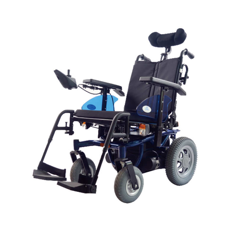
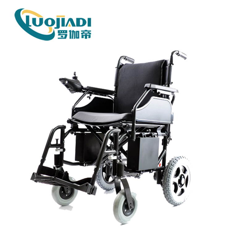
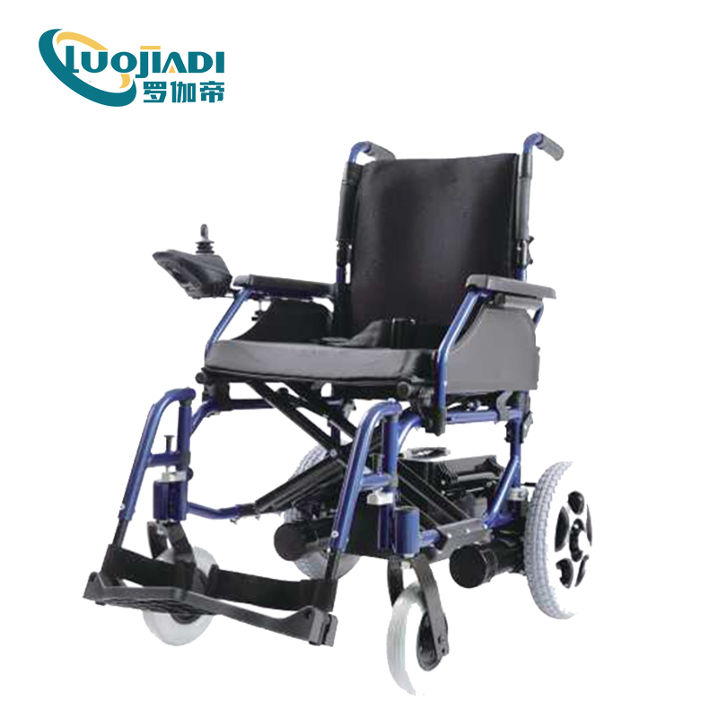
The fundamental difference from traditional electric scooters, battery scooters, bicycles and other transportation tools is that electric wheelchairs have intelligent operating controllers. According to the different operation methods, there are rocker-type controllers, and controllers controlled by various switches such as the head or blowing system. The latter is mainly suitable for severely disabled people with upper and lower limbs. Nowadays, electric wheelchairs have become an indispensable means of transportation for the elderly and the disabled. It is suitable for a wide range of objects. As long as the user has a clear consciousness and normal cognitive abilities, the use of electric wheelchairs is a good choice, but it requires a certain amount of space for activities.
power wheelchair,hospital use,homecare product,surgucal equipment Shanghai Rocatti Biotechnology Co.,Ltd , https://www.ljdmedicals.com


